Regional Anesthesia in the Emergency Department

USF Residents Participate in Dr. Derr’s Ultrasound Guided Regional Anesthesia Workshop
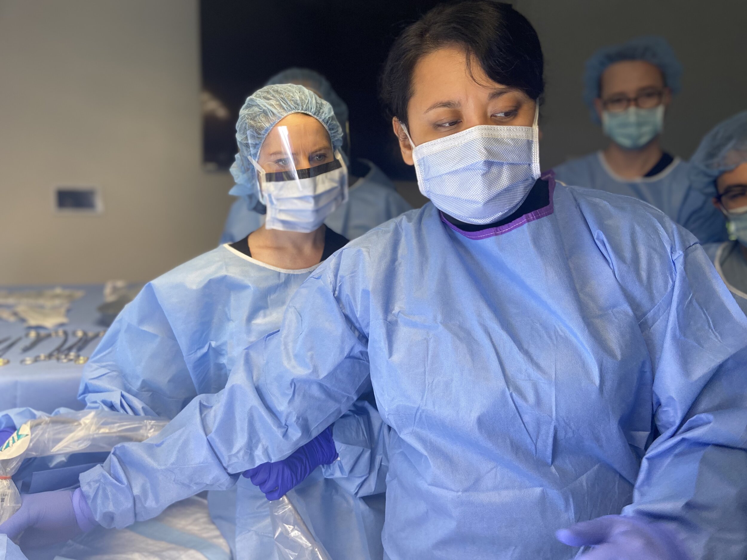
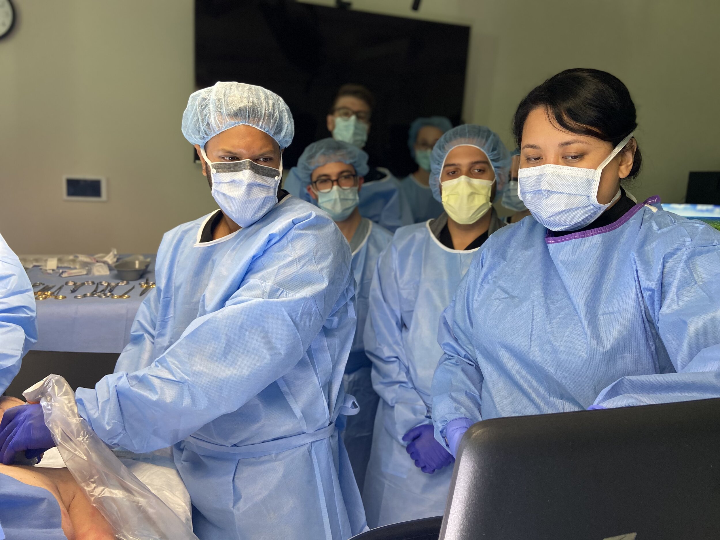
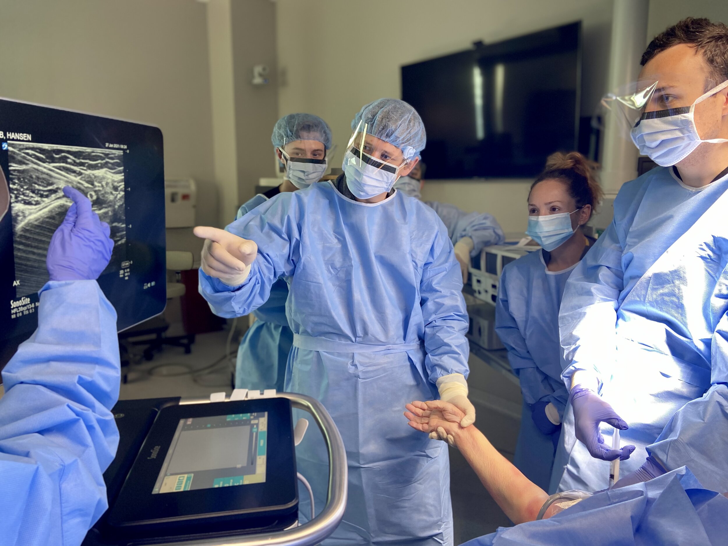
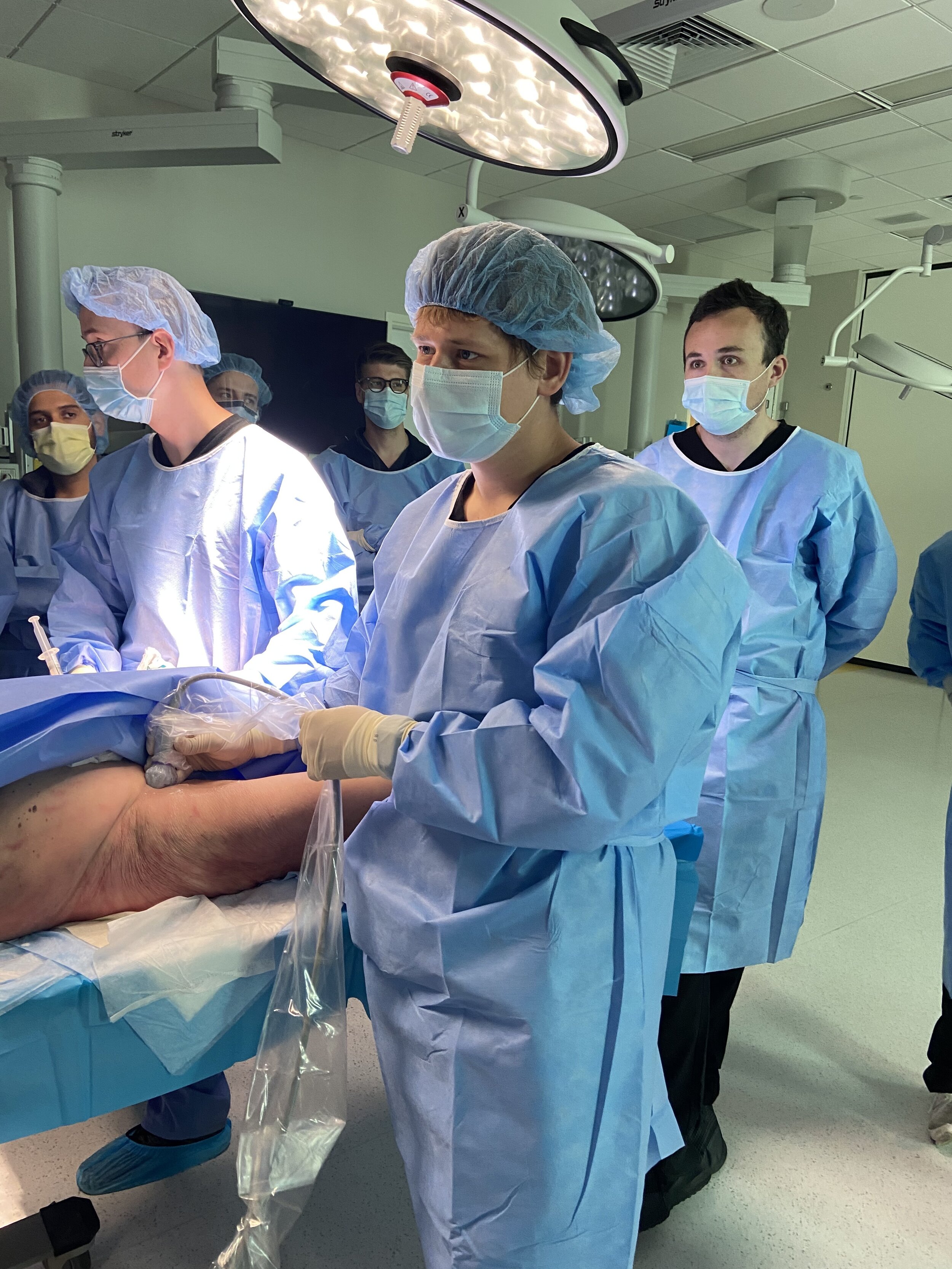
Our interns recently spent the day in the cadaver lab training in ultrasound-guided regional anesthesia (UGRA). Regional anesthesia can be an extremely effective way to manage pain and minimize opioids. Despite the potential benefits, not all emergency medicine residencies offer training in regional anesthesia. Wilson et al. reported that 30% of programs do not offer any UGRA training. Thanks to Dr. Derr & our Ultrasound Faculty, USF offers training in regional anesthesia to our residents. During last year’s workshop, I learned how to successfully perform a posterior tibial nerve block. Over the past year, I’ve had the opportunity to use this technique multiple times and I love it!
Posterior Tibial Block
The reason I love this block: The bottom of the foot is sensitive and impossible to effectively achieve adequate pain control without using this block. This block is perfect for complex lacerations or foreign bodies in the sole of the foot. The first time I used this block it was for a patient who had injured their foot on coral. The repair required a lot of irrigation, foreign body removal, and complex wound repair.
Indications: Injuries and procedures involving the sole of the foot.
Complications: This procedure is generally uncomplicated. All nerve blocks may result in bleeding, infection, and damage to nearby structures. Neuritis is usually temporary. Other major complications that have been reported include intra-arterial injection resulting in toxicity or vasospasm.
“The landmarks for this block, the medial malleolus and the posterior tibial artery, make it tempting to do this technique blindly. Unfortunately, the blind technique doesn’t work very well. I’ve had better success using the ultrasound-guided approach. ”

How to Perform a Posterior Tibial Block
1. Position the patient and clean the medial ankle with chloraprep.
2. Place the linear probe just posterior to the medial malleolus.
3. Locate the neurovascular bundle. The posterior tibial nerve is located posterior to the tibial artery and veins. Nerves have a distinct hyperechoic honeycomb appearance.
4. Insert a 22g needle using an in-plane or out-of-plane approach. If using an in-plane approach, the needle should enter just anterior to the Achilles tendon. In the out-of-plane approach, the needle is inserted inferior and perpendicular to the probe.
5. Anesthetize the skin and advance the needle until it is in close proximity of the nerve, but you do not want to inject into the nerve.
6. Aspirate prior to injection to ensure the needle is not in a vessel. Inject 5cc of local anesthetic in close proximity to the nerve so that the nerve is bathed in the local anesthetic.
If the patient complains of pain or shocking sensation during injection, stop injecting and withdrawal the needle slightly to avoid injecting into the nerve.
“The posterior tibial nerve block can be performed using an in-plane or out-of-plane approach. This is one of the few nerve blocks where the operator may prefer to enter with the needle out of plane, because the posterior tibial artery and the Achilles tendon make an in-plane approach technically difficult. ”

The vessels and the nerve are shown above. Note the honeycomb appearance of the nerve. Click the photo to watch a great video from 5 min Sono.
Check out this video from 5 min Sono:
https://www.coreultrasound.com/posterior-tibial-nerve-block/
In my next blog entry, we will discuss local anesthetic systemic toxicity (LAST). LAST is something we should think about every time we use local anesthetic and especially if we begin to incorporate UGRA. Though there is very little risk of LAST associated with the posterior tibial block, toxicity can be a real risk with some of the other nerve blocks requiring larger volumes. UGRA can be safely performed when clinicians understand LAST and how to prevent it.
References
Wilson CL, Chung K, Fong T. Challenges and Variations in Emergency Medicine Residency Training of Ultrasound-guided Regional Anesthesia Techniques. AEM Educ Train. 2017 Feb 18;1(2):158-164. doi: 10.1002/aet2.10014. PMID: 30051027; PMCID: PMC6001815.
https://www.coreultrasound.com/posterior-tibial-nerve-block/
https://www.acep.org/sonoguide/posterior_tibial_nerve_block.html
About the Author
Dr. Okonkwo is the Assistant Program Director at USF. She attended medical school at Indiana University and completed her emergency medicine residency training at Carolinas Medical Center in Charlotte, North Carolina. This post was reviewed by Dr. Charlotte Derr, Program Director and Ultrasound Fellowship Director at USF Emergency Medicine.