
Awake and Facilitated Intubations
Let’s Define it!
Awake intubation is a technique in which a patient is intubated without the use of paralytics or sedatives. Intubation is instead achieved through the use of local anesthesia. The goal of this is to preserve the patient’s protective airway reflexes and respiratory drive.
Facilitated intubation is almost identical to an awake intubation but uses the adjunct of light sedation, often Versed or Ketamine. Airway reflexes are still preserved in this method. This is often used in patients who may be too anxious/non-cooperative with typical awake intubation.
Indications
Any patient who you anticipate a difficult airway for!
While there is not a well validated predictor of difficult airway some patient’s you may want to consider reaching for this are
Patient with head or neck pathology (malignancy, surgery, radiation)
Patient with reduced mouth opening
Those with limited neck extension (especially elderly pts)
The morbidly obese patient
Those with a history of sleep apnea
Patient with significant cardio/pulmonary risk factors (those who will desaturate quickly and may not give you time for a second attempt if paralyzed)
Those who you suspected impending respiratory compromise (Think anaphylaxis and smoke inhalation pts)
Contraindications
Patient Refusal
The immediately crashing patient.
Techniques/Essential Equipment
Rare and less familiar procedures, such as these, can give rise to some anxiety and hesitation. However, when breaking down this procedure into ints individual steps, it becomes much more manageable.
Pre-oxygenation
While you are gathering supplies, put that time spent to good use and start preoxygenating your patient.
Often you can place a Non-rebreather mask (NRB) or NRB + Nasal Cannula. For patients with higher oxygen requirements, you can consider NIPPV (Bipap or Hi-flow)
Once you are ready to intubate switch the patient to nasal cannula for passive oxygenation during the procedure without compromise of access.
Dry them out
For the best views and for proper efficacy of the topical anesthetic it is best to minimize the presence of oral secretions by choosing a medication to minimize secretions.
Typical medications used are:
0.2 mg glycopyrrolate
0.01 mg/kg of atropine
This takes about 15 minutes to work, so consider doing this step while setting up your pre-oxygenation.
Topicalize
This is likely the step most unfamiliar to us in the ED but let’s break it down.
Get 4% Lidocaine, placed in nebulizer mask, run at 5L – this will anesthetize the oral cavity.
Obtain an atomizer device and continue with atomized lidocaine. At this point the oral cavity should be numb and you can direct the mist further back in the oropharynx
Allow the patient to gargle viscous lidocaine.

If you can’t find an atomizer device or it has to “come from central” you can get crafty!
Items you need: 20g IV catheter, O2 tubing, 3-way stopcock, 10 cc syringe with lidocaine. Attach as shown here and run the O2 at around 5L.
Sedate
If you do not think your patient will tolerate being awake for intubation now is the time for sedation
Options for sedation
Ketamine (given in 20mg increments)
Low dose Versed
Ketofol
Remember to go light on sedation, the key is to keep those airway reflexes intact.
You may want to consider soft restraints as the sedated patient may reflexively pull at the ETT.
Intubate
Switch to Nasal Cannula to give yourself room to work and your patient passive O2
Obtain your airway box/cart and have adjuncts ready just like a normal intubation.
Make sure to have paralytic & sedation ready for post intubation
Do not forget to have your suction ready.
Position the patient and the bed for optimal success.
Ideally auditory canal at level of sternal notch
Some patients may need to head of bed elevated depending on pathology.
Head elevation to 30 degrees can reduce risk of passive regurgitation.
Proceed with Orotracheal intubation in method of your choosing.
Direct – Can be useful potentially bloody airways or those centers without video laryngoscopy.
Video-Laryngoscopy (VL) – Can help obtain great views, especially if patient requires positioning sub-optimal to operator or potential blood or contaminant in airway.
Fiber-optic – Can help in patients with potential for structural abnormalities (stenosis, masses, etc) and those with limited mouth opening which may impede visualization with DL or VL. May not be the best choice for airways with heavy secretions or blood.
DL/VL + Bougie
Confirm placement
Most recommend at minimum two-point check (visualization through cords and end-tidal) which is something we do for every intubation anyway. So, treat this just like your typical RSI, confirm in the same ways you usually do and get the confirmatory CXR
Post-Intubation
Provide sedation and paralytic of your choice based on clinical scenario.
Optimize your ventilation settings based on clinical scenario.
Now a Case!
HPI & Presentation
Patient presents via EMS with reported epistaxis from left nostril refractory to prolonged pressure on unknown blood thinner.
Vital signs are grossly stable, patient is protecting airway with intermittent cough.
Patient states nose started bleeding several hours prior to arrival to ED and happened spontaneously.
Patient has had similar episode in past but is a poor historian in general with no family to assist.
Patient is slightly agitated, and EMS was unable to obtain IV access in route to ED.
Paramedic is assisting holding pressure and patient is sitting straight up in stretcher.
Initial interventions
ENT kit, TXA, rhino rockets and suction are all called for and set up in room.
Constant pressure is held for 10-15 minutes while setting up adjunct treatment options and airway supplies are collected.
Bleeding fails to be controlled with direct pressure and sitting patient forward.
TXA and afrin are added to direct pressure without success.
Patient is unable to tolerate passing of rocket and becomes extremely agitated despite use of pain dose ketamine.
Patient’s daughter to bedside in attempt to calm patient to tolerate rockets with re-attempt
Patient still unable to tolerate rocket with daughter bedside.
The Facilitated Look
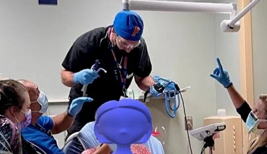
Physicians decide to perform a facilitated look with dissociative ketamine. In prepping for this approach the physicians called for backup methods to bedside including RSI medications, direct laryngoscope, supraglottic airway, bougie, and surgical airway supplies. Prior to pushing dissociative ketamine, the physicians palpate the neck to ensure they are prepared for a surgical airway if needed.
Dissociative dose ketamine given and video-assisted intubation performed. An assistant performed suctioning allowing for the physician to get a clear view of the cords.
Once the cords were successfully visualized, rocuronium was administered. The patient was kept in the upright position and oxygenated via non-rebreather mask until the cords were paralyzed. The physician was able to pass a 7.5 ET tube using video-assisted intubation with rigid stylet and double suction set up.
Tube placement confirmed with ETCO2, auscultation, and CXR.
Post-intubation and ED Course
Post intubation care initiated with copious suctioning and OG tube placed.
Large amounts of clots continued to collect in oropharynx despite bilateral rockets placed and inflated.
Patient’s mouth packed with tube gauze while awaiting emergent ENT consult.
Patient’s hemodynamics remained stable, one unit of type and crossed blood given with suspected acute blood loss and marginal Hgb.
Patient’s daughter provided physician much needed hug for handling of care.
Ultimately, ENT was able to control the patient’s bleeding and the patient made a full recovery.
References
https://associationofanaesthetists-publications.onlinelibrary.wiley.com/doi/full/10.1111/anae.14904
http://www.emdocs.net/awake-endotracheal-intubation/
https://emupdates.com/awake-intubation-a-very-brief-guide/
http://pemsource.org/2016/11/29/improvised-mucosal-atomizer-device/
about the authors
Dr. Ken Dumas is a PGY-1 at USF Emergency Medicine. He completed medical school at the University of Central Florida in Orlando, FL. Following graduation he plans to pursue an EMS Fellowship. Dr. Jullian Beau is an Attending Physician at Tampa General. He also serves as the Course Director for the USF EM teaching elective. He completed medical school at Charles E. Schmidt College of Medicine (FAU) in Boca Raton. He went on to complete his emergency medicine residency at Emory University in Atlanta, GA.